As time went on, of course, I began to make sense of what I was seeing and to relate the nerves I was dissecting to the segmental arrangement of the spinal cord. But it took me some time to feel reasonably confident that I understood what I was doing.
When it comes to the brain similar problems arise. Brain anatomy is intimidating for most of us and it's all too easy to get lost in the welter of details. What you want at the outset is just a basic idea of how the brain is organised and where its main components are located.
There are plenty of books and internet resources which supply this kind of information, but even these can seem rather daunting the first time you encounter them. I've come to believe that it's best to start, not with the mature brain, but by going back literally to the beginning and seeing how the brain develops over time to take the form it does.
That means starting with embryology. You might think that this would be
an 'advanced' subject, and of course the embryology of the nervous
system is indeed very complex, but the essentials are easy to grasp.
This approach works for me and I hope it may work for you too. If it
doesn't, forget it and take another route to understanding.
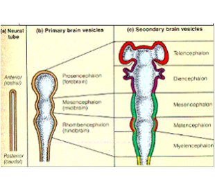
In its simplest state the central nervous system is just a closed tube (the neural tube). The front end will become the brain and the rest will form the spinal cord (Figure 1a). This is true of the embryo, and it also describes how the nervous system has developed in the course of evolution. You probably began your biological studies by dissecting 'lower' animals such as worms and the dogfish, so you will be familiar with these more 'primitive' forms of the nervous system. To a certain extent embryology 'recapitulates' evolution.
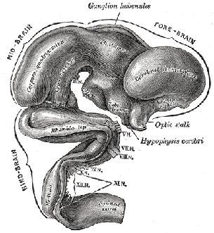
Very soon the front of the neural tube acquires three bulges (the primary brain vesicles). These are destined to become the forebrain, midbrain and hindbrain (Figure 1b). The neural tube now looks like those segmented party balloons that you may have blown up as a child. So far, of course, there is no resemblance to the adult human brain, but have a little patience.
Next (Figure 1c), the forebrain acquires some side bulges (secondary brain
vesicles). The two at the front are the future cerebral
hemispheres—the biggest and most obvious parts of the adult brain as we look at it
from the outside. (There are two smaller cup-shaped bulges just behind
these, which will become the eyes, but we don't need to bother about
those here.)
At this point we can see several things.
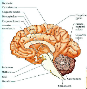
(a) The neural tube has now been folded over, which brings the floor of the forebrain into contact with the floor of the hindbrain (Figure 2). This will cause various structures in the mature brain to lie closer together than they would be if the doubling-over of the neural tube hadn't happened.
(b) The hindbrain has acquired some new bulges, which will become the cerebellum.
(c) The hollow tube is the forerunner of the ventricles of the brain.
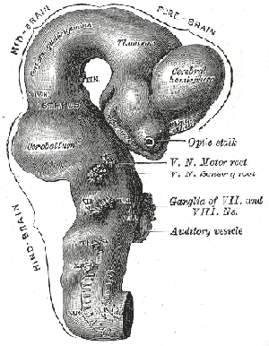
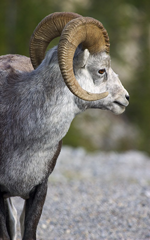
The folding already apparent in Figure 2 has now increased. Notice also that the developing cerebral hemispheres are beginning to curl round, making them appear rather like the horns of a ram (Figure 4).
By now the cerebral hemispheres have expanded and curled further and are
beginning to look more like a brain as we usually think of it. The two
lobes of the cerebellum can also be seen although they are still quite
small.
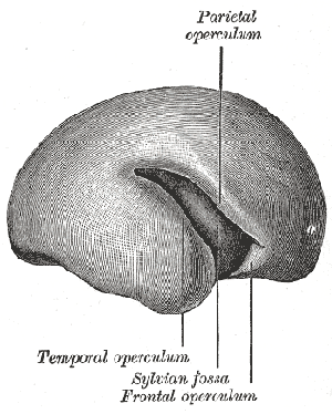
In shape, this reminds me of a boxing glove (Figure 7).

Of course, there are two cerebral hemispheres, so two boxing gloves,
side by side (Figure 8).

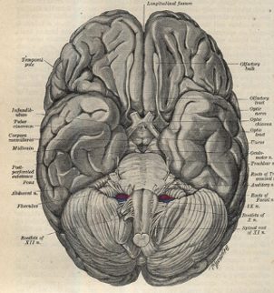
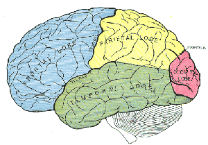
In terms of the boxing glove analogy, the fingertips correspond to the
frontal lobe, the back of the glove is the parietal lobe, the wrist is
the occipital lobe, and the thumb is the temporal lobe. The hollow
interior of the glove corresponds to the ventricles and the leather on
the outside to the cerebral cortex.
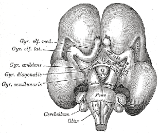
(b) The hippocampus is formed by a folding up of the inner margin of the temporal lobe. This gives it a 'Swiss roll' appearance in cross-section. Note that it is, therefore, part of the cerebral cortex (Figure 11).
(c) The cingulate gyrus is a layer of cortex on the inside of the glove,
adjacent to the little finger (Figure 12).